


Orthodontic treatment (braces) is a common and effective way to improve both your bite and your smile. To ensure treatment is accurate and successful, your orthodontist needs a comprehensive understanding of your teeth, jaws, and facial structure. That’s why taking the appropriate dental X-rays is an essential part of the initial examination and treatment planning process.
Each type of X-ray serves a specific purpose, helping the orthodontist diagnose problems, monitor progress, and develop the most effective treatment plan. It’s also important to note that not every patient needs every type of X-ray. In most cases, only one or a few types are recommended based on the orthodontist’s clinical assessment. Therefore, it’s best to have an examination first and only undergo the imaging your orthodontist prescribes, which can also help reduce unnecessary costs.
So, what types of dental X-rays are commonly used before orthodontic treatment? Let’s take a closer look.
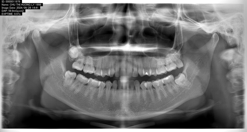
1. Panoramic X-ray (Panoramic Radiograph)
A panoramic X-ray provides a broad view of the entire mouth, including the upper and lower jaws, all teeth, the temporomandibular joints (TMJs), wisdom teeth, and impacted teeth.
This type of X-ray also helps orthodontists identify abnormalities such as impacted teeth, jaw cysts, bone lesions, or other conditions that may affect treatment. It offers an overall assessment of both the teeth and jawbone before an orthodontic treatment plan is developed.

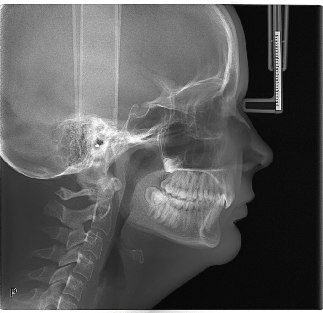
2. Lateral Cephalometric X-ray (Cephalometric Radiograph)
A lateral cephalometric X-ray is a side-view image of the skull that shows the relationship between the teeth, jaws, and facial bones.
This X-ray allows the orthodontist to measure various angles and distances between skeletal and dental structures, helping determine whether bite problems—such as protrusion (overbite) or underbite—are caused primarily by the teeth or by the underlying jawbone. Based on this information, the orthodontist can plan precise tooth movements while maintaining facial harmony.
This is considered one of the most essential X-rays for nearly every orthodontic patient.
3. Periapical X-ray
A periapical X-ray focuses on one or a few teeth, providing detailed images of the entire tooth, including the root and the surrounding bone.
It helps detect conditions such as root infections, bone loss, and other abnormalities around the tooth root. Periapical X-rays are also valuable for evaluating root length, which is particularly important before and during orthodontic treatment, as teeth with shorter roots may be at a higher risk of root resorption during tooth movement.
This type of X-ray is typically prescribed when the orthodontist suspects a problem affecting specific teeth.
4. Bitewing X-ray
A bitewing X-ray is taken while the patient gently bites on a small film or digital sensor placed between the upper and lower teeth. It provides a clear view of the contact areas between the back teeth.
This X-ray is especially useful for detecting cavities between teeth—areas that are often difficult to examine visually—as well as evaluating bone levels between adjacent teeth.
In orthodontics, bitewing X-rays are not routinely required for every patient. They are generally recommended when there is concern about tooth decay or periodontal (gum) disease.
5. Cone Beam Computed Tomography (CBCT)
Cone Beam Computed Tomography (CBCT) is a three-dimensional imaging technique that provides highly detailed images of the teeth, jawbones, and surrounding anatomical structures, including nerves.
CBCT scans allow orthodontists to evaluate jawbone anatomy in greater detail, assess impacted teeth, and examine complex wisdom teeth. This imaging method is particularly valuable in challenging cases involving impacted teeth, planned tooth extractions, severe skeletal discrepancies, or situations where orthodontic treatment is combined with jaw surgery.
Why Are X-rays So Important Before Orthodontic Treatment?
Taking the appropriate dental X-rays before—and sometimes during—orthodontic treatment is essential. They enable the orthodontist to accurately evaluate the condition of your teeth and jawbones, identify potential problems, and create a treatment plan that is safe, effective, and tailored to your individual needs.
Each type of X-ray provides unique information, and together they give a complete picture that supports successful orthodontic care.
If you’re considering braces or are already undergoing orthodontic treatment, be sure to follow your orthodontist’s recommendations regarding dental imaging. Having the right X-rays at the right time is an important step toward achieving the best possible treatment outcome.